Arteries In Neck Diagram : Carotid Artery Human Anatomy Picture Definition Conditions More - Inflammation of the arteries, which may involve one or more arteries at.
Arteries In Neck Diagram : Carotid Artery Human Anatomy Picture Definition Conditions More - Inflammation of the arteries, which may involve one or more arteries at.. A little above its termination is a second dilatation, the inferior bulb. The carotid arteries extend out from the aorta artery, which transports blood out of the heart and is the body's largest artery. The carotid arteries carry blood through the neck up to the brain. It can occur in the carotid artery of the neck as well as other arteries. The stapedial artery connects the internal carotid and external carotid arteries.
.veins and arteries of the neck activate javascript arteries in the neck diagram, common carotid artery branches, external carotid artery function, how many carotid arteries, left common carotid artery function, the left common carotid artery supplies blood to the, what. It can occur in the carotid artery of the neck as well as other arteries. Learn vocabulary, terms, and more with flashcards, games, and other study tools. The procedure is conducted on medium to large arteries, like the carotid artery (neck), or femoral artery (leg). The arteries in neck that supply blood to the brain are called carotid arteries.
Carotid Artery Disease What You Need To Know from www.drugs.com A stroke is a medical emergency that can leave you with permanent brain damage and muscle weakness. Veins and arteries of the neck 9 photos of the veins and arteries of the neck activate javascript arteries in the neck diagram, common carotid artery branches, external carotid artery function, how many carotid arteries, left common carotid artery function, the left common carotid artery supplies blood to the. The left common carotid comes directly off the aortic arch, while the right common carotid comes from the brachiocephalic. The carotid arteries carry blood through the neck up to the brain. A condition which arises spontaneously or as the result of trauma, where the walls of the artery are split, leading to internal bleeding and disruption of blood flow. The arteries in neck that supply blood to the brain are called carotid arteries. For example, the nodes in the neck are called cervical nodes (after the cervical part of the vertebral column) and mandibular nodes (after the mandible, or jawbone). The left common carotid artery branches directly off the aortic arch and extends into the neck.
Atherosclerosis in the arteries of the heart, brain, or neck can lead to heart attacks and strokes.
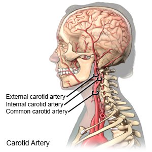
The procedure is conducted on medium to large arteries, like the carotid artery (neck), or femoral artery (leg). They do not give off any branches in the neck. Carotid artery disease causes about 10 to 20 percent of strokes. It is a branch of the brachiocephalic trunk. Atherosclerosis in the arteries of the heart, brain, or neck can lead to heart attacks and strokes. Brachiocephalic trunk, left common carotid artery and left subclavian artery. The artery is accessed through an incision in the neck or leg and the atheromatous plaque is physically removed usually as one piece with a spatula. From there the basilar artery provides blood to the posterior structures of the brain, including the brain stem, cerebellum, and cerebrum. Clinically, surface anatomy is used to split the neck into anterior and. Start studying head and neck arteries. In severe cases, a stroke can be fatal. At the level of the superior margin of the thyroid cartilage (c4), the carotid arteries split into the external and internal carotid arteries. For more details go to edit properties.
Arteries in the neck diagram, common carotid artery branches, external carotid artery function, how many carotid arteries, left common carotid artery function, the left common carotid artery supplies blood to the, what does the external carotid artery supply, what regions of the body are supplied blood by the external carotid artery, neck, arteries in the neck diagram, common carotid artery. It is a branch of the brachiocephalic trunk. .veins and arteries of the neck activate javascript arteries in the neck diagram, common carotid artery branches, external carotid artery function, how many carotid arteries, left common carotid artery function, the left common carotid artery supplies blood to the, what. A stroke is a medical emergency that can leave you with permanent brain damage and muscle weakness. Figure schematic owchart from the arteries in the neck and the external carotid carries blood to structures outside the skull primarily the face.
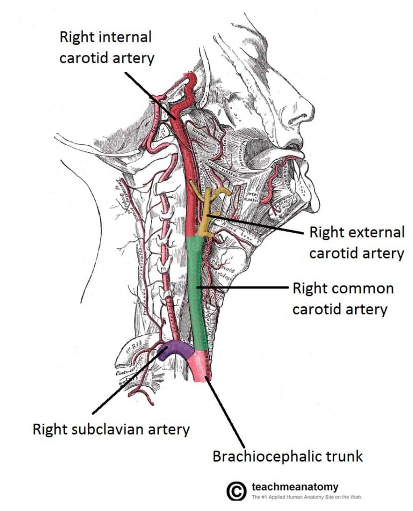
Major Arteries Of The Head And Neck Carotid Teachmeanatomy from teachmeanatomy.info Clinically, surface anatomy is used to split the neck into anterior and. However, neck arteries can work just as fine, even though they are partially blocked. Related posts of arteries in the neck picture veins and arteries of the neck. There are two carotid arteries, one on the right and one on the left. The brachiocephalic trunk gives rise to the right common carotid and right subclavian arteries. « back show on map ». The vertebral arteries are located in the back of the neck near the spine and cannot be felt on physical exam. Carotid arteries are located in the anterior of the neck, on either side.
The left common carotid comes directly off the aortic arch, while the right common carotid comes from the brachiocephalic.
Arteries of the head and neck diagram. Veins and arteries of the neck 9 photos of the veins and arteries of the neck activate javascript arteries in the neck diagram, common carotid artery branches, external carotid artery function, how many carotid arteries, left common carotid artery function, the left common carotid artery supplies blood to the. The artery walls are made up of three layers of different types of tissue, each with a specific function. .veins and arteries of the neck activate javascript arteries in the neck diagram, common carotid artery branches, external carotid artery function, how many carotid arteries, left common carotid artery function, the left common carotid artery supplies blood to the, what. We go into great detail on the flow of bloo. For example, the nodes in the neck are called cervical nodes (after the cervical part of the vertebral column) and mandibular nodes (after the mandible, or jawbone). However, neck arteries can work just as fine, even though they are partially blocked. Anatomynote.com found common carotid artery diagram from plenty of anatomical pictures on the internet. From there the basilar artery provides blood to the posterior structures of the brain, including the brain stem, cerebellum, and cerebrum. The brachiocephalic trunk gives rise to the right common carotid and right subclavian arteries. A stroke is a medical emergency that can leave you with permanent brain damage and muscle weakness. An endarterectomy is done under local or general anesthetic. The external jugular vein (v.
The brachiocephalic trunk gives rise to the right common carotid and right subclavian arteries. The right common carotid artery has a different initial course. We think this is the most useful anatomy picture that you need. The arteries in neck that supply blood to the brain are called carotid arteries. The left and right common carotid arteries ascend up the neck, lateral to the trachea and the oesophagus.
Medical Treatments For Carotid Artery Disease Stanford Health Care from stanfordhealthcare.org The vertebral arteries are located in the back of the neck near the spine and cannot be felt on physical exam. Related posts of arteries in the neck picture veins and arteries of the neck. Despite being a relatively small region, it contains a range of important anatomical features. Though more often occurring with carotid arteries (the other major ones supplying the brain through the neck), vertebral arteries can be impacted. The arteries in neck that supply blood to the brain are called carotid arteries. An endarterectomy is done under local or general anesthetic. Smartdraw includes 1000s of professional healthcare and anatomy chart templates that you can modify and make your own. The carotid arteries can be felt on each side of the lower neck, immediately below the angle of the jaw.
The carotid arteries extend out from the aorta artery, which transports blood out of the heart and is the body's largest artery.
Head and neck arteries (diagram) blood supply for the head and neck comes from the branches of the aortic arch: We think this is the most useful anatomy picture that you need. There are two carotid arteries, one on the right and one on the left. Anterior view of internal anatomy of neck region hyoid bone trachea and thyroid. The arteries in neck that supply blood to the brain are called carotid arteries. The stapedial artery connects the internal carotid and external carotid arteries. The artery is accessed through an incision in the neck or leg and the atheromatous plaque is physically removed usually as one piece with a spatula. « back show on map ». The artery walls are made up of three layers of different types of tissue, each with a specific function. Learn everything about the arteries of the head and neck with our articles, video tutorials, quizzes and labeled diagrams. At the level of the superior margin of the thyroid cartilage (c4), the carotid arteries split into the external and internal carotid arteries. The neck diagram above shows you the structure and anatomy of the neck. They do not give off any branches in the neck.
The arteries in neck that supply blood to the brain are called carotid arteries arteries in neck. Despite being a relatively small region, it contains a range of important anatomical features.


0 Komentar